Macular degeneration can lead to extensive vision loss which occurs in the central area of your field of vision. Your central point of sight may become blurry or you may develop a blind spot. Macular degeneration typically occurs during the aging process. Although it has genetic roots, lifestyle factors can increase the severity or progression of this disease.
Dry macular degeneration is the most common type and is a slow progressing form of the disease caused by thinning and degeneration of the center of the retina, called the macula. This can develop into the rapidly progressing wet macular degeneration, caused by blood or fluid leaking from abnormal blood vessels into the macula.
Early Detection Can Slow the Progression of Macular Degeneration
In order to detect macular degeneration, your doctor may administer certain tests. Some of the following tests are rarely used, but your doctor will perform one or more of these tests to make a proper diagnosis. If a diagnosis of macular degeneration is made, you may be put on certain medications or be asked to make certain lifestyle changes in order to slow the progression of the disease to help prevent central area vision loss.
1. Peeping at a Visual Acuity Test to Check for Changes in Your Peepers
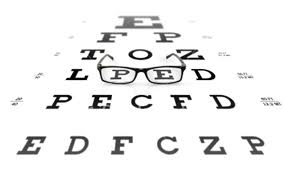
This is the same test used to determine if you need to get glasses or contacts. It is typically a white chart with black letters of varying sizes used to determine how small of letters you can see clearly at 20 feet of distance. A change in your vision since your last examination may simply mean that you need vision correcting lenses. However, it might also mean that you have an eye condition that requires further testing.
2. Spying the Amsler Grid Test to Detect Eye and Vision Problems
The Amsler Grid is a patterned testing device that can be used at the doctor’s office or at home in between doctor’s visits to monitor any progression of macular degeneration. This testing grid consists of a white grid with crossing lines printed on a black background. When you follow the instructions, all of the lines will remain straight and visible unless you are developing macular degeneration. In that case, the lines will appear wavy or distorted, or might even disappear from the field of vision.
3. Examining the Back of the Eye to Check for Abnormalities in the Retina
The eyes will be dilated so that the doctor can use a special instrument to examine the retina, which is located in the back of the eye. The doctor will check for any abnormalities. One point of concern is to check for the development of yellow deposits under the retina that are called drusen. These form more frequently in those with dru macular degeneration. The doctor will also check for symptoms of wet macular degeneration, which includes the observation of fluid or blood that has leaked into the macula.
4. Glancing the Surface of the Retina with a Scanning Laser Ophthalmoscope
A Scanning Laser Ophthalmoscope is a special tool that uses a bright beam of light to get detailed images of the surface of the retina, blood vessels, choroid, and optic disk. This test may be used in connection with a basic retinal exam since the eyes are already dilated. A doctor might choose this option if results from a basic dilated retinal examination are not clear or when the results are inconclusive. The Scanning Laser Ophthalmoscope can also be used for Microperimetry to determine macular sensitivity and macular fixation pattern.
5. Scanning the Layers of the Retina with Optical Coherence Tomography
The different layers of the retina can be examined with cross-sectional imaging. These layers can be examined for thickness, distortion, or swelling. Thinning areas can also be detected. This test is a non invasive way to not only detect problems in the retina, but also to assess the effectiveness of any treatment options that are being used to control macular degeneration and to slow its progression.
6. Picturing the Retina with Fundus Photography to Compare Results over Time
While the pupils are dilated, the doctor may decide to use Fundus photography to take pictures of the retina, the macula, and the optic nerve. A special camera is used that allows for documentation of changes to the parts of the eye over a period of time to determine the progression of any disease. This is typically used in connection with other tests including retina tests such as Scanning Laser Ophthalmoscope and Optical Coherence Tomography.
7. Pressing the Cornea with Tonometry to Test Intraocular Pressure
You doctor might decide it is necessary to check the pressure inside of the eyes. This is called tonometry. This test is typically used to check for glaucoma and certain other eyes conditions. Depending on the type of tonometry used, your eyes might be numbed prior to testing by the use of special eye drops. Pressure can be measured with certain tools that actually touch the surface of your eye, or may involve non-contact methods, such as using a puff of air to measure the pressure. The air puff method is not very accurate, but it does not require the use of numbing drops to be placed in the eyes prior to testing.
8. Dyeing for Wet Macular Degeneration with Fluorescein Angiogram
If the doctor suspects wet macular degeneration, a Fluorescein Angiogram might be ordered. For this test, the eyes will be dilated and pictures of the retina will be taken if they haven’t already. Then a contrast dye will be injected into a vein in the arm where it will travel to the eyes. The contrast dye makes the blood appear fluorescent. Pictures will again be taken as the dye will show the location of any leaking blood vessels. This test might be repeated after a period of time to see if the disease is progressing.
9. Getting into Your Genes with Genetic Macula Risk Testing
The Macula Risk test is a genetic test used to predict how macular degeneration will progress in the individual being tested. It is most frequently used in those already diagnosed with the disease to help the doctor develop the best treatment plan. It is a simple test that uses a small brush to swab the inside of the cheek for cells. This swab is then sent to the lab for results that can be read by the doctor. This test is not always necessary for the doctor to come up with an effective treatment plan to slow progression of the disease.
10.Measuring Macular Pigment Density with QuantifEye to Detect Thickness of Protection from Light Damage
Macular pigment is a protective barrier inside the eye that also aids in vision acuity, color vision, contrast, and glare control. Light can damage eyes over time, and this barrier can help reduce the risk of that damage. However, this protection thins over time, allowing more light to enter the sensitive parts of the eye. QuantifEye is a special tool that uses heterochromatic flicker photometry technology to detect the thickness of this macular pigment barrier, thus showing if you have an increased risk of developing macular degeneration.
